Showing posts with label Heart. Show all posts

Wednesday, May 26, 2021

How much take Salt, sodium and potassium in healthy diet

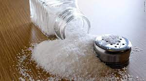
Most people consume too much sodium through salt (corresponding to consuming an average of 9–12 g of salt per day) and not enough potassium (less than 3.5 g). High sodium intake and insufficient potassium intake contribute to high blood pressure, which in turn increases the risk of heart disease and stroke.

How-much-take-Salt-sodium-and-potassium-in-healthy-diet
how much take Salt

Reducing salt intake to the recommended level of less than 5 g per day could prevent 1.7 million deaths each year.

People are often unaware of the amount of salt they consume. In many countries, most salt  comes from processed foods (e.g. ready meals; processed meats such as bacon, ham and salami; cheese; and salty snacks) or from foods consumed frequently in large amounts (e.g. bread). Salt is also added to foods during cooking (e.g. bouillon, stock cubes, soy sauce and fish sauce) or at the point of consumption (e.g. table salt).

How-much-take-Salt-sodium-and-potassium-in-healthy-diet
Salt

Salt intake can be reduced by:

  • limiting the amount of salt and high-sodium condiments (e.g. soy sauce, fish sauce and bouillon) when cooking and preparing foods;
  • not having salt or high-sodium sauces on the table;
  • limiting the consumption of salty snacks; and
  • choosing products with lower sodium content.

Some food manufacturers are reformulating recipes to reduce the sodium content of their products, and people should be encouraged to check nutrition labels to see how much sodium is in a product before purchasing or consuming it.

Potassium can mitigate the negative effects of elevated sodium consumption on blood pressure. Intake of potassium can be increased by consuming fresh fruit and vegetables.

How-much-take-Salt-sodium-and-potassium-in-healthy-diet
Salt


Friday, May 14, 2021

Heart murmurs

Just as the ECG gives important information about the electrical operation of the heart, heart sounds provide valuable information about its mechanical operation. A heart murmurs is an abnormal sound that consists of a flow noise that is heard before, between, or after the lubb-dupp or that may mask the normal heart sounds. 

Although some heart murmurs are 'innocent' meaning they do not suggest a heart problem, most often a murmur indicates a valve disorder. 

Among the valvular abnormalities that may contribute to murmurs are mitral stenosis (narrowing of the mitral valve by scar formation or a congenital defect), mitral insufficiency (back flow or regulation of blood from the left ventricle into the left atrium due to a damaged mitral valve or ruptured chordae tendineae), aortic stenosis (narrowing of the aortic semilunar valve), and aortic insufficiency (backflow of blood from the aorta into the left ventricle). 

Another cause of a heart murmurs is mitral valve prolapse (MVP), an inherited disorder in which a portion of a mitral valve is pushed back too far (prolapsed) during ventricular contraction. 

Although a small volume of blood may flow back into the left atrium during ventricular systole, mitral valve prolapse often does not pose a serous threat. 

In fact, it is found in up to 10% of otherwise healthy young persons. 

Wednesday, October 9, 2019

Hypertension Types and Causes

Hypertension-type-and-causes
Hypertension
Hypertension is also known as high blood pressure. Pressure exerted by the blood against wall of blood vessels. Blood pressure rises to about 110-120 mm hg during ventricle contraction (systole) and fall to about 80-70 mm hg during ventricle relaxation (diastole).  
 
Hypertension can lead to severe complications and increase the risk o heart disease, stroke and death.
Long term high blood pressure is a major risk factor for heart failure, coronary artery disease, stroke, vision loss, chronic kidney disease and peripheral vascular diseases.

Two type of hypertension

Primary hypertension or essential hypertension       
 
This type of hypertension results from a complex interaction of genes and environmental factors. 

Secondary hypertension  

It results from an identifiable cause. Renal disease is the most common cause of secondary hypertension. 

Cause of Hypertension

Blood pressure rises with ageing and the risk of becoming hypertensive in later life is considerable.
  • Excessive drinking alcohol
  • Renin angiotension system
  • Smoking high salt intake
  • Obesity
  • Depression
  • Excessive eating liquorice
  • Headaches
  • Vertigo
  • Anxiety
  • Excessive sweating
  • Buzzing in the ear
  • Treatment
  • Stop drinking alcohol
  • Stop smoking
  • Lack salt intake
  • Exercise
Take anti hypertensive drugs – captopril, enalapril, losartan, propranolol, atenolol, verapamil.

Heart Function and Mechanism

Heart-Function-and-Mechanism
Heart
Heart is conical and hollow organ. 

Location of heart

The heart lies in the thorax between the lungs behind the sternum.
Two third of the heart is on the left side

Long - 12 Cm
wide  - 9 cm
thick - 6Cm
in adult female, heart weight – 250gm
in adult male, heart weight – 300gm

Pericardium

The pericardium membrane is surrounds and protect the heart. 

The pericardium consist of two main parts:

Fibrous pericardium 

The superficial fibrous pericardium is composed of tough inelastic dense connective tissue.

Serous pericardium 

The deeper serous pericardium is thinner or more delicate membrane that forms double layer around the heart.

The outer parietal layer of the serous pericardium is fused to the fibrous pericardium.

The inner visceral layer of the pericardium also called epicardium.

Between the parietal and visceral layer of the serous pericardium is a thinner film of lubricating serous fluid.

Layer of heart wall

The wall of the heart consist of three layer
  • Epicardium (External layer)
  • Myocaridum (middle layer)
  • Endocardium (inner layer)

Chamber of the heart

The heart has four chambers.

The two superior receiving chambers are the arteria and interior pumping chambers are ventricles.

Right atrium

The right artrium receive blood three veins: 
  1. The superior vana cava 
  2. Inferior vana cava 
  3. Coronary sinus
Veins always carry blood toward the heart.
Arteries always take blood away from the heart.
Blood passes from the right artrium into the right ventricle through a valve that is called the tricuspid valve. Right atrium thickness around 2-3mm. it is also known as right atrioventricular valve.

Right ventricle

It thickness is 4-5mm. the right ventricle is seprated from the left ventricle by a thinner wall, called the interventricular septum. Blood passes from the right ventricle through the pulmonary valve in to a large artery called the pulmonary artery and carries blood into the lungs.

Left atrium

Its thickness 2-3mm. it receives blood from the lungs through pulmonary vein.
Blood passed from the left atrium into the left ventricle through the bicuspid valve (mitral valve or semilunar valve)

Left ventricle

Its thickness is 10-15mm. blood passes from the left ventricle through the aortic valve in to the ascending aorta. Some of blood in to the aorta flows into the coronary artery which carry blood to the heart wall. Blood passes into the arch of the aorta.

Valve of heart:

Tricuspid valve
 
That valve present in between right artrium and right ventricle.

Pulmonary valve

That valve present between right ventricle and pulmonary artery.

Bicuspid valve

That valve is present between left atrium and left ventricle.

Aortic valve or aortic semilunar valve

That valve is present in between the left ventricle and aorta.

Blood circulation

It is classified into four parts:
  1. Systemic circulation
  2. Pulmonary Circulation
  3. Coronary circulation
  4. Portal circulation

Systemic circulation

It is the circulation involving blood supply to all parts of the body except lungs. This circulation starts from aorta (which carries oxygenated blood from left ventricle).

Pulmonary Circulation

It is the part of the circulation involving the purification of blood in lungs. The pulmonary artery carries deoxygenated blood from right ventricles to lungs, where oxygen molecules are bind with hemoglobin and blood became oxygenated.

Coronary circulation

The circulation involve to the heart cells and tissues.
 

Portal circulation

Portal vein carries blood (that has circulated blood in stomach, intestine and pancreas).

Function of heart

The heart is act as a pump. It maintains a constant circulation of blood throughout the body.

Mechanism of heart


Deoxygenated blood comes from cells and tissues in right atrium through superior vena cava, inferior vena cava and coronary sinus. 

De-oxygenated blood fill in right atrium and tricuspid valve is open. Blood transfer into right atrium.

Deoxygenated blood goes lungs through pulmonary artery, where deoxygenated blood is change into oxygenated blood (oxygen bind to haemoglobin molecules of blood). Oxygenated blood is transfer from lungs to left atrium through pulmonary vein. Bicuspid valve is open and blood transfer to left ventricle. 

Oxygenated blood is send to all body cells and tissues by arch of arota. Coronary artery supply oxygenated blood to heart tissues.